

In our exploration of iron metabolism and overload, we uncovered the pivotal role of the liver as the primary reservoir for iron storage. But the question remains: How can we accurately measure the amount of iron stored in the liver without resorting to invasive procedures?
Traditionally, liver iron concentration has been evaluated through liver biopsy, a procedure that entails extracting a small liver tissue sample for chemical analysis. While effective, this method is invasive, uncomfortable, and not conducive to repeated measurements. Moreover, the procedure carries inherent risks and limitations, making it less than ideal for routine clinical use.
Thankfully, advancements in medical technology have paved the way for non-invasive alternatives, capitalising on the magnetic properties of iron to provide accurate assessments of liver iron concentration. Two notable techniques that have emerged in this regard are superconducting Quantum interference device-based magnetic susceptometry (SQUID) and magnetic resonance imaging (MRI).
SQUID, a physics-based technique, harnesses the magnetic properties of iron to measure liver iron concentration. This method involves applying a strong magnetic field to the liver while detecting changes in signal intensity as the liver moves away from the detector. While SQUID has demonstrated efficacy in quantifying liver iron concentration, its widespread adoption is hindered by the scarcity of SQUID machines, which are limited in availability and accessibility.
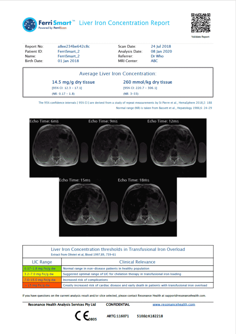
On the other hand, MRI offers a more accessible and versatile approach to assessing liver iron concentration. MRI scanners are ubiquitous in hospitals and radiology clinics, making them a convenient option for non-invasive evaluation. By exploiting the magnetic properties of iron-containing particles within the liver, MRI can generate quantitative images that reflect liver iron concentration. This technique, known as FerriScan, utilizes water molecules in the tissue as antennas to detect magnetic fields emitted by iron-containing particles. Through sophisticated mathematical algorithms, FerriScan produces detailed maps of liver iron distribution, enabling clinicians to assess iron concentration with a precision better than biopsy.
The advent of MRI technology has revolutionised the field of liver iron quantification, offering a safer, more comfortable, and more accessible alternative to traditional biopsy techniques. With MRI, patients can undergo assessments without the need for invasive procedures or recovery periods, enhancing the overall patient experience and compliance with diagnostic protocols.
In the diagnostic pathway for hereditary hemochromatosis and other iron-related disorders, MRI plays a pivotal role in confirming the presence of iron overload and guiding appropriate treatment decisions. When genetic tests do not conclusively identify the presence of hereditary haemochromatosis, FerriScan provides a definitive means of assessing iron status, enabling clinicians to tailor treatment strategies to individual patient needs effectively.
Moreover, FerriScan enables clinicians to monitor changes in liver iron concentration over time, providing valuable insights into the efficacy of treatment interventions and disease progression. By tracking iron levels longitudinally, healthcare providers can optimize patient care and outcomes, ensuring that individuals with iron-related disorders receive timely and appropriate interventions to mitigate the risks associated with iron overload.
In conclusion, innovative approaches to assessing liver iron concentration, such as MRI-based techniques like FerriScan, represent a significant advancement in the field of iron metabolism and overload. By offering a non-invasive, accurate, and accessible means of quantifying liver iron concentration, FerriScan enhances our ability to diagnose, monitor, and manage iron-related disorders effectively, ultimately improving patient care and outcomes.
How can Resonance Health help?
Our products FerriScan®, FerriSmart® and LiverSmart® can analyse LIC with high reliability. These methods use MRI imaging, so they are non-invasive and painless. Contact us to know more.